


Reviewed Date: 09/15/2026
✔ Medically Reviewed By: Naveed Javied, PT
Disclaimer: Always seek the advice of your physician, physical therapist, or other qualified healthcare provider with any questions you may have regarding a medical condition, chronic pain, or before starting any new rehabilitation, exercise, or nutritional program. Never disregard professional medical advice or delay in seeking it because of something you have read in this article.
Table of Contents
Close your eyes and touch your nose. You didn’t miss, did you? You didn’t need a mirror to guide your index finger to its target. That invisible, effortless coordination relies entirely on a sensory system most people take for granted until it suddenly stops working.
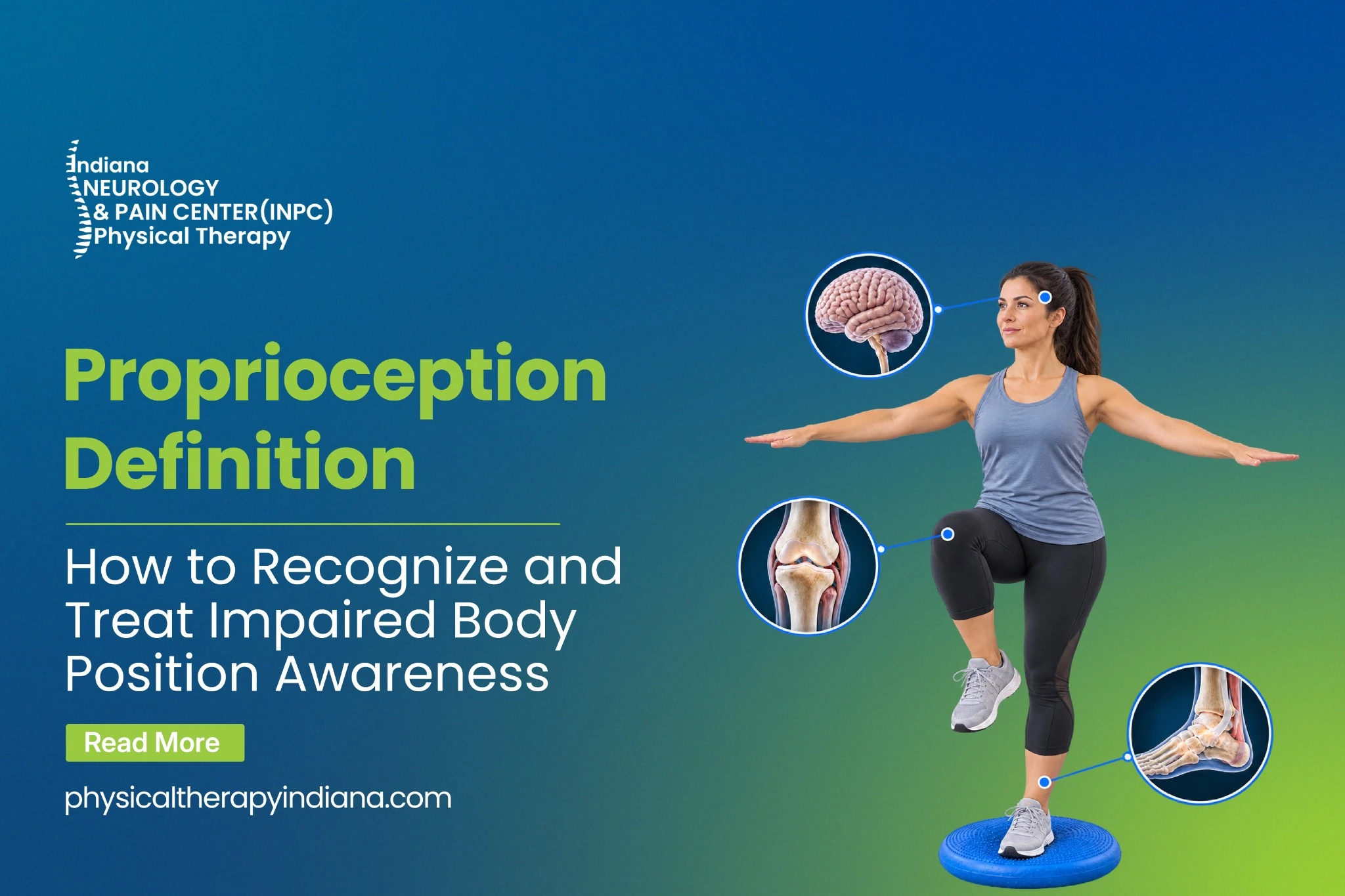
When physical therapists talk about restoring movement, establishing a solid proprioception definition usually comes first. Derived from the Latin word proprius—meaning “one’s own”—this is your body’s innate ability to sense its own position, movement, and force in space. It is the absolute biological foundation of body position awareness. It lets a typist hit the right keys without looking at their hands. It keeps a runner upright when they hit an unexpected patch of gravel.
The Core Concept: Decoding the Terminology
In medical quizzes and rehabilitation assessments, students and patients alike often ask: Which of the following best defines proprioception?
The truest definition is that it operates as a continuous, high-speed neuromuscular feedback loop. It isn’t just one nerve or one specific brain region. Instead, millions of microscopic sensory receptors buried inside your muscles, tendons, and joint capsules act as biological GPS trackers. They constantly stream data regarding muscle stretch and joint angles through the peripheral nerves, up the spinal cord, and into the brain. The brain crunches this massive wave of data in milliseconds to coordinate fluid, purposeful movement.
People often confuse it with kinesthesia. They are incredibly close siblings, but they aren’t twins. While kinesthesia strictly deals with sensing behavioral movement, the proprioceptive system is broader. It encompasses both dynamic movement and static posture. It tells your brain exactly where your limbs are, even when you are standing perfectly still.
The Neurology: Types of Proprioception
To really grasp how the system functions, you have to look at the wiring. The human nervous system splits this spatial tracking into two distinct pathways. These different types of proprioception dictate how we react to the physical world around us.
First, there is conscious proprioception. These signals travel up the dorsal column-medial lemniscus pathway straight into the cerebrum. This allows a person to actively recognize where a limb is placed. If a doctor moves your ankle while you are blindfolded, you can verbally describe exactly how the joint is bent.
Second, we have unconscious proprioception. These electrical impulses bypass conscious thought entirely, traveling via the spinocerebellar tracts directly into the cerebellum. This pathway handles rapid-fire, automatic postural corrections. When you trip on an uneven sidewalk and your foot instantly snaps forward to catch your fall, you didn’t think about it. Your unconscious system saved you.
The Hardware: How Your Body Tracks Itself
How does this biological GPS actually collect data? It relies on mechanoreceptors. These tiny anatomical structures detect mechanical changes in tissue and translate them into electrical nerve impulses.
Table 1: The Primary Receptors of the Proprioceptive System
| Receptor Type | Where It Lives | What It Does | Triggered By |
|---|---|---|---|
| Muscle Spindles | Buried deep inside the belly of skeletal muscles. | Acts as the body’s stretch-detector to prevent over-lengthening. | Changes in muscle length and how fast that stretch occurs. |
| Golgi Tendon Organs | Sitting right at the junction where muscle fibers meet tendons. | Monitors tension to prevent muscles from tearing themselves apart under heavy loads. | Extreme muscle tension and mechanical force. |
| Joint Kinesthetic Receptors | Tucked inside joint capsules and surrounding ligaments. | Provides critical feedback on joint angles and acceleration. | Extreme joint rotation and structural stress. |
Whenever you move, these receptors fire off simultaneously. Your brain synthesizes this incoming sensory data, cross-references it with what your eyes see and what your inner ear (vestibular system) feels, and issues outgoing motor commands.
Everyday Proprioception Examples
Because this processing happens behind the scenes, it can be hard to visualize. So, what does a healthy system actually look like? Consider a few common proprioception examples:
- Navigating in the dark: Walking from your bed to the hallway switch without visually tracking the floor. Your brain knows exactly how far to stride.
- Applying the brakes: Driving a car and knowing exactly how much pressure your foot needs to apply to the brake pedal without ever looking down.
- Climbing stairs: Lifting your foot to the exact height of the next step.
- Holding a paper cup: Gripping a full paper coffee cup firmly enough so it doesn’t slip, but gently enough so it doesn’t crush in your hand.
When the Signal Drops: Impaired Proprioception
What happens when the communication lines get cut? A patient develops impaired proprioception, and suddenly, the brain is flying blind.
The causes range from severe neurological conditions to everyday sports injuries. Degenerative diseases like multiple sclerosis or Parkinson’s literally strip away the neural pathways. Peripheral neuropathy, often driven by diabetes, suffocates the nerve endings in the feet and hands. The signal never even makes it to the spinal cord.
But the most common culprit in the clinic is structural joint damage.
Think about a severely sprained ankle or a torn ACL. When you rip a ligament, you aren’t just tearing collagen fibers. You are ripping out the joint kinesthetic receptors living inside that tissue. The hardware is physically destroyed. This is exactly why an ankle might feel “wobbly” or give out months after the swelling is gone. The tissue healed. The neurological software did not.
Table 2: Healthy Function vs. Sensory Deficits
| Daily Activity | Intact Sensory Function | Signs of a Sensory Deficit |
|---|---|---|
| Walking & Gait | Fluid, even steps; looking ahead at the environment. | “Stomping” the feet to feel the ground; constantly staring at the floor. |
| Static Balance | Stable posture, even when closing the eyes. | Swaying heavily or immediately falling when visual cues are removed. |
| Fine Motor Control | Effortlessly picking up keys or coins. | Clumsiness, dropping objects, or crushing fragile items by gripping too hard. |
| Spatial Navigation | Walking through doorways without issue. | Frequently bumping into walls, doorframes, or misjudging distances. |
Clinical Assessments and Rehabilitation
To figure out how bad the damage is, clinicians use highly specific tests.
The Romberg test is a classic baseline: you stand with your feet together and shut your eyes. If you immediately start to fall, it proves your body was relying entirely on vision to stay upright. To test specific joints, therapists use Joint Position Sense (JPS) testing. They move a patient’s limb to a specific angle, bring it back, and ask the patient to replicate that exact angle while blindfolded. Sports physical therapists might also use the Y-Balance test, asking an athlete to balance on one leg while reaching outward in different directions to spot hidden asymmetries.
The good news? You can fix it. Neuroplasticity allows the brain to literally rewire itself. Through targeted physical therapy, patients can completely rebuild their body position awareness.
Rehabilitation isn’t just about lifting heavy weights. It’s about sensory confusion and controlled instability.
Therapists will usually start with a basic single-leg stance on a flat, solid floor. Once the brain masters that, they pull the rug out. They introduce unstable surfaces—BOSU balls, foam pads, and balance boards. The wobble board forces the ankles and knees to make hundreds of rapid micro-adjustments. The muscle spindles and joint receptors are forced to wake up and fire rapidly.
Next, therapists remove the visual system. Doing a single-leg squat with your eyes closed forces the somatosensory system to do 100% of the heavy lifting. Finally, reactive training comes into play. A therapist might randomly toss a ball at a patient balancing on one leg, or apply unexpected pushes (perturbations) to train automatic joint stabilization. The goal is to make the nervous system bulletproof before the patient steps back onto a basketball court or simply navigates an icy sidewalk.
Conclusion

Naveed Javied, PT
Physical Therapist
Naveed Javied is a highly skilled physical therapist specializing in orthopedic rehabilitation. He earned his Doctor of Physical Therapy from the University of Montana, building upon his Master of Physical Therapy from Quinnipiac College. With extensive clinical experience, including specialized orthopedic care at Oakwood Annapolis Hospital, Javied focuses on helping patients effectively manage pain and restore their mobility.
Frequently Asked Questions
Think of it as your body’s secret inner eyes. Even when your real eyes are closed, your muscles have tiny little sensors that tell your brain exactly what your arms and legs are doing.
They are incredibly close, but there is a line drawn between them. Proprioception includes your sense of balance and static posture—knowing your arm is bent even when you aren’t moving. Kinesthesia is strictly about the behavioral awareness of movement.
It depends entirely on the root cause. If you lost your balance because of a sprained ankle or a knee surgery, yes. Physical therapy can absolutely retrain those local receptors and get you back to 100%. However, if the loss is driven by a progressive neurological disease (like MS), a complete “cure” isn’t likely. Instead, physical therapy shifts toward symptom management, helping the patient develop compensatory strategies to stay mobile and safe.
Alcohol acts as a heavy depressant on the central nervous system, specifically targeting the cerebellum. Since the cerebellum handles unconscious proprioception, alcohol effectively scrambles the signal. Your brain processes the location of your feet a fraction of a second too late. This delayed feedback loop causes the slurred speech, swaying, and stumbling we associate with intoxication. Field sobriety tests are literally just testing your proprioceptive pathways.
References
https://pmc.ncbi.nlm.nih.gov/articles/PMC10483674/
https://pubmed.ncbi.nlm.nih.gov/9204262/